health and akita

veterinary

What are the risks of buying a non-pedigree Akita? (from untested parents)
"Little Leia's Story"
The following story comes from a US mailing list:

“I don't know much about the place where I was born. It was narrow and dark and the man never played with us.
I still remember my mother and her soft fur, but she was often sick and very thin. She had little milk for me and my brothers and sisters. Most of them died suddenly. When they took me away from my mother, I was so scared and so sad. My baby teeth had barely come out and I still needed my mother so much. Poor mother, she felt very bad herself.
People said that now they finally wanted the money and that my sister's and my squealing were getting on their nerves.
So one day we were loaded into a crate and taken away. We hugged each other and felt both of us shaking, unconscious with fear. Nobody came to comfort us. All these strange sounds and smells.
We are in the "Pet Shop" - a store where there are many different animals. Some meow, others squeak, some whistle. We also hear other puppies whining. My sister and I press tightly together in a tight cage. Sometimes people come and watch us, often quite small people who look very friendly, as if they wanted to play with us. We spend day after day in our little cage. Sometimes someone catches us and lifts us up to look at them. Some are friendly and pet us, others are rude and hurt us. We often hear them say, "oh they're cute, I want one," but then people leave again.
Last night my sister died. I laid my head on her soft fur and felt the life drain from her thin body. When they took her out of the cage in the morning, they said that she was sick and that I should be sold cheaper so that I would go soon. No one pays attention to my silent cries when my little sister is thrown out.
Today my family came and bought me!
Now everything will be fine! These are very nice people who actually decided on me. They have good food and a beautiful bowl, and this little girl carries me very gently in her arms. Her dad and mom say I am a very sweet and brave dog. My name is Lea now. I can even kiss my new family, it's great. They warmly teach me what I can and cannot do, they take care of me, they give me delicious food and lots and lots of love. I want nothing more than to please these wonderful people, and nothing is more beautiful than running around and playing with a little girl.
First visit to the vet. It was a strange place, I was shaking. I got several injections. My best friend, a little girl, holds me gently and tells me it will be okay, so I relax. The vet must be saying something sad to my lovely people, because they look quite devastated. I hear something about severe defects, E dysplasia and heart disease. He talks about wild breeders and how my parents were never tested. I didn't understand any of it, but it was terrible to see my family so sad.
I'm six months old now. My peers are wild and strong, but every move causes terrible pain. The pains never stop. Plus, I immediately run out of breath whenever I want to have some fun with my little girl. I would love to be a strong dog, but I just can't do it... My father and mother talk about me. I see them all so sad and it breaks my heart. In the meantime, I go to the vet often and it always ends with "genetic" and "nothing can be done". I want to play outside in the warm sun with my family, I want to run and jump. I can't…
Last night was worse than any other. I couldn't even get up to drink and I just screamed in pain... They carried me to the car. Everyone is crying. They're so weird, what's going on? Have I been naughty? Are they angry at me? No, no, they caress me so gently. Oh, if only these pains would stop! I can't even lick the tears off the little girl's face, but at least I can reach her hand.
The vet's table is cold. I'm afraid. People cry into my fur, I feel how much they love me. With some effort I manage to lick their hand. The vet takes his time today and is very cordial and I feel a little less pain. The little girl holds me very gently, a slight twinge… Thank God, the pain disappears. I feel deep peace and gratitude...
Dream: I see my mother, my brothers and sisters in a big green meadow. They call to me that there is no pain there, only peace and happiness.
So I say goodbye to my human family the only way I can: with a gentle wag and a light sniff. I wanted to spend many happy years with you, it shouldn't have happened. Instead, I only caused you a lot of worry. I'm sorry, I was just a commodity after all.
"Lea"
©1999 J. Ellis (translation by I. Bach-Żelewska)

DYSPLASIA – causes – what the breeder can do and what the owner should do!
Dysplasia, simply put, is a deformation of the hip joint. The acetabulum is so flat that it does not fully cover the head of the femur. Dysplasia most often affects dog breeds that grow very intensively during puppyhood.

Based on research, the Swedes determined the probability of dysplasia in puppies from various matings:
both parents are free from dysplasia: 24%- 37.5% (If all grandparents are free from dysplasia, the probability is only 8.7 %!)
one parent with dysplasia: 44.7% – 50%
both parents with dysplasia: 84.1% to even 93 %!
In the USA, the reliability of X-rays at different ages was examined: 12 months - 68.9 %, 18 months - 82.7 %, and 24 months - even 95.4 %. This means that it is possible that a 1-year-old dog with mild dysplasia may have healthy joints at the age of 2 (due to ongoing development). So if we have a dog of high value with slight dysplasia, we should not use it, but we should not withdraw it from breeding - let's repeat the tests at the age of 2 years.

The international rules for combating dysplasia in dogs are:
Pair dogs with normal hip joints;
Normal dogs should come from normal parents and grandparents;
Normal parents should produce 75% healthy offspring;
Partners with the highest % of born normal offspring (at least 75%) should be selected for mating;
For breeding, you should choose female dogs with perfectly shaped hip joints - better than those of their parents.
Of course, dysplasia also involves environmental factors, i.e. the influence of the environment:
poor conditions for raising puppies, overfeeding them, poor diet (lack of balanced food, which practically only ready-made food and appropriate supplements can provide to a large breed puppy). On the so-called Home-made food (which some people call table scraps - with a lot of spices and other things not recommended for a dog) will not raise a healthy large breed puppy (even if you bought it from a reputable breeder - from both parents free of dysplasia).
At this point, I appeal to you: if you cannot afford to properly feed a large dog (and especially in the first year the amounts are really significant), please do not buy a puppy and do not waste its health and life. Or postpone the purchase for a while and save to have financial reserves for the time of its growth, because once you buy a dog, you cannot postpone its proper feeding until later. Every bag of cheap food from the Supermarket or cheap Chapi or Pedigree you buy will take its revenge very quickly. If you save on good food (because the lady from the pet store recommended a cheap one), you will actually soon spend many times the saved amount on a veterinarian, and often without any effect. So if you cannot afford proper (unfortunately expensive) feeding of a large breed dog, please take a small dog from a shelter (its feeding will be much cheaper) and sometimes there are really beautiful gems there that really need a home! Why, driven only by the snobbish desire to show off to your friends, should you buy a beautiful, large dog that you cannot really afford to keep? You will waste it because you will regret buying high-quality food and vitamins that the breeder says and you will try to replace it with home-made food and an egg yolk or a bit of cottage cheese added from time to time. This dog, which would be wasted elsewhere at your place, would be a happy and healthy dog if well cared for and properly fed, and the breeder would not suffer to see you destroy a wonderfully promising dog, which he took care of from the day he was born until he was handed over to you.

Another cause of dysplasia is the inappropriate "use" of the puppy, i.e. lack of exercise or excess exercise - overloading the growing joints, as well as the type of exercise (e.g. climbing stairs to the 4th floor, running on slippery floors, where the paws spread apart without having a stable surface, a young puppy running on a bike, playing with too large and heavy dogs), poorly organized bed.
Puppies should receive high-quality food containing appropriate amounts of collagen substances, balanced in terms of minerals. Foods that cause obesity increase the risk of disease. If hip dysplasia is suspected, the only sure diagnosis can be made by X-ray. A reliable assessment of the joint can be made at the age of 12-18 months. The first symptoms of dysplasia can be observed at many 6 months. A dysplastic puppy may then limp and have difficulty jumping, climbing stairs and standing up. During the course of the disease, the joint becomes more damaged and the symptoms become more severe. So if for some reason you are concerned about your puppy's movement, you can check it. It is recommended that this inspection and a possible X-ray be performed by a veterinarian who is authorized to enter the dysplasia result in the pedigree, because only then can we be sure that he is not only a true professional but also a person with high ethics and that he will not deceive naive owners. a puppy, for example, for surgery on a healthy dog just to earn several hundred zlotys.
Attention! If, unfortunately, you have an X-ray taken by a veterinarian other than an authorized one, be sure to request the photo (the photo is your property and you paid for it) - and consult the diagnosis with an authorized veterinarian, so as not to unnecessarily cut up a healthy dog just because a poor veterinarian took a bad X-ray photo or read it incorrectly or, worse still, deliberately told us that we had a sick dog just to earn a few hundred zlotys.) If the doctor refuses to provide you with the photos and insists on surgery, I advise you to stay away from such clinics. Because if you don't have a photo, you will never prove to him that he not only cheated you out of a large sum of money, but what's worse, he unnecessarily exposed your dog to enormous pain and mutilation, which without his intervention would have been healthy until the end of its days - and will remain crippled forever. result of unnecessary surgery.
For example, trimming the adductors does not change the impairment of the hip joints (if it actually exists) and only temporarily reduces the pain. However, this procedure completely prevents the placement of endoprostheses in the future. International veterinary recommendations regarding the resection of femoral heads (in dogs with a target weight above 20 kg) clearly state: "absolutely not recommended!" Such thoughtless resection in a large breed dog, which is in the phase of intensive growth, may result in impairment of the second acetabulum (previously healthy) and deformation of the femur.
* Only photos of adult dogs taken for the purpose of entering the dysplasia result in the pedigree (by an authorized doctor) must remain in the clinic and are archived there (this is a requirement to obtain an entry in the pedigree). The remaining photos are always the property of the dog owner who pays for them.
ANDJewish Bach-Żelewska
* * *
ATTENTION! the following two articles were placed here with the consent of: Marzena Kaca-Bibrich (original source: www.z-palacu-cesarza.j.com.pl)
Dr. vet. med. Stefan Jung (translation: W.Ziemecki, consultation: dr.hab J.Siembieda)

"Does nutrition influence joint diseases?"

Degenerative joint diseases such as dysplasia and osteoporosis are not uncommon in dogs. Despite the dog's outstanding predispositions, factors related to the dog's upbringing and nutrition may influence the occurrence and development of the disease. The so-called glucosaminoglycans are of particular importance in this respect.
Dogs of all ages can suffer from joint diseases. Even under the Animal Protection Act, the person responsible for the dog should do everything (with reasonable effort) to protect it from these diseases, which are chronically degenerative, painful and limit the lifespan. Breeders of these so-called negatively predisposed breeds who attach great importance to their good name and dog competitors who want to enjoy their dogs for as long as possible should seriously address the relationship between the development of degenerative joint diseases and the measures that can prevent it.
Two joint diseases have been prominent in medical practice for years, which mainly affect large dogs and which are similar in origin: hip dysplasia (HD) and osteochondroisis dissecans (OCD). The main topic of this article should be the development of HD, as it is more common than OCD.
Anatomical basics
Before we discuss the disease and its development, we must discuss several processes without which a complete understanding of the disease would be impossible.
Cartilage is a flexible, multi-layered tissue that consists of cartilage cells (chondrocytes), connecting fibrin (collagen fibers) and ground substance (matrix). The calcification zone contains few cells and is embedded directly on the bone surface and is attached to it. This bond is intended to prevent the cartilage from separating from the bone under load. For this purpose, cells have receptors for vitamin D3 during calcification, thanks to which calcium not only enriches the binding, but can also control it itself
The middle zone (attrition zone, transition zone) contains a large amount of cells, connective tissue and matrix. This zone acts as a water cushion that can absorb and distribute heavy loads that affect the cartilage surface. The proximal zone lying closest to the joint gap has a specific connective tissue structure, such as fibers parallel to the surface, which limits the cartilage tissue in relation to the joint gap.
Functionally, a joint is a flexible connection of two bones. The joint consists of two bone epiphyses with epiphyseal cartilage and partly an internal cartilaginous binding apparatus that holds and stabilizes both ends of the bones together from the inside. The empty spaces of the joint contain the so-called joint lubricant (synovia), which ensures good sliding of the cartilage surfaces and supplies valuable nutrients to the cartilage cells. In large joints (e.g. hip) we find "Hyalina" cartilage, which has a very high water content; large joints are always surrounded by a joint capsule; muscles and tendons stabilize the joint only from the outside. "Fibrocartilage" is found especially in the intervertebral discs of the spine.
Joint diseases
Hip dysplasia (HD)
Canine HD is a disease that affects especially large, heavy breeds, but also increasingly medium breeds. For many years, breeders, breed associations and veterinarians have tried to work together to reduce the increasing incidence of this disease. In these efforts, multiple solutions were formulated with more or less substantive courage. The fact is that HD, as before, is the most common disease in certain breeds.
While previously it was believed that HD was a genetically determined problem, today it turned out that the formation of HD is conditioned by at least one more thematic complex: the dog's (poor) nutrition! A method of movement that is not very physiological (e.g. sports involving jumping over obstacles) seems to play a minor role. There are controversial debates as to what percentage a given thematic complex influences the formation of HD. The authors Hazewinkel and Wiegand give the genetic factor 20 to 50 percent, so other factors may account for 50 to 80 percent.
When we talk about the development of a disease, we must always take into account two concepts that interact: predisposition and condition. By predisposition we understand the sum of inclinations inherited from parents, they are given by nature. By fitness we mean the sum of all acquired features.
However, poor condition (e.g. due to insufficient nutrition) does not necessarily have to destroy good predispositions in the long term. However, it is a known fact that animals with a certain predisposition (inherited from one or both parents) can develop predispositional properties (e.g. HD) more quickly than animals without these predispositions.
The complex issue of hereditary traits can be dealt with relatively easily if the breeder and buyer make sure that, ideally, both parents are free from dysplasia or are classified with a low degree of dysplasia. This classification is performed using x-rays. In addition to radiology, HD examinations should always include the assessment of joint elasticity.
HD is manifested anatomically by the fact that the joint socket and the head of the femur are incongruent, i.e. they do not fit each other exactly. When assessing the X-ray, the adaptation of the articular surfaces of the pelvic acetabulum and the head of the thigh bone is determined and the so-called "Norberg angle" is measured.
The second complex of topics (poor) nutrition, according to today's knowledge, largely contributes to the development of HD. Most authors mainly see three nutritional mistakes:
overfeeding (food energy and material)
excess calcium and/or
too high vitamin D3 content in the food
With this type of nutritional errors, good predispositions may not prevent the development of HD.

Adult dogs that eat a daily dose of high-calorie food are most often overweight, sluggish and later suffer from organ diseases or metabolic diseases caused by excess weight. Puppies behave differently: the hip joint of a newborn dog is first made of cartilage. The calcification process that produces the finished joint, bone elongation, and weight development are inherently interrelated. Feeding high in energy leads to rapid weight gain, which begins to damage the joints and their elastic cartilage during puberty, even when puppies "look healthy." As a result, HD does not develop spontaneously, but only during the first 15 months of life.
Another factor in the formation of HD is excessive enrichment with calcium or calcium compounds (lime for puppies). Adding calcium to puppies is only recommended when feeding them "home-made" or when they show symptoms of rickets. Ready-made puppy foods are usually well-balanced. It's not just about the absolute calcium content of the food; the possible content of vitamin D3 should also be taken into account, which increases the body's calcium intake. Too high levels of calcium in the blood result in an accelerated bone remodeling process, which most often ends in defective mineralization (thin, brittle bones). In addition, we should also mention the influence of the changed metabolism of the acid base caused by nutrition or the excessive presence of particular compounds, e.g. sodium, potassium, chloride.
In the case of HD in a young dog, two different symptoms of the disease are described: younger dogs, up to 15 months of age, show a sudden reluctance to run and pain in the hind limbs, get up with difficulty and move reluctantly. The musculature of the hind limbs is poorly developed and cannot keep up with the growth of the bones, so the entire load must be borne by the large joints, which overloads the cartilage. Although still elusive X-ray, cartilage changes begin in the form of microcracks (small scratches) and the first scars, which are the result of overload.
Older dogs with X-ray evidence of HD most often develop limping after heavy loads. HD manifests itself with more or less twisted ankle joints, a swinging gait and partial sparing of the affected limb in a standing position. In some animals, the defective formation of the ankle joint leads to chronic, high cartilage overload with increased signs of wear (fissures and scars), which manifest themselves in inflammation and pain and further increase the mismatch.
In both cases, the joint cartilage is not able to cope with the load, it develops first small, then increasingly larger scratches (cracks), which become inflamed and heal. Since these areas of inflammation are of low functional value, they tear further, resulting in loss of substances. Calcium salts often accumulate in these inflamed areas, leading to the formation of so-called "noses"; these ossified noses are convex and harder than the opposite surface of the cartilage, as a result of which they destroy it.
Osteochondrosis dissecans (OCD):
The development of OCD is multifactorial and very similar to HD, so we will only describe the picture of the disease. OCD most often occurs in the elbow joint of large breeds. It almost exclusively affects young dogs.
It affects animals aged four to eight months, and limping occurs spontaneously or after trauma. The cause is microcracks that become deeper. Unlike HD, however, a larger piece of cartilage is detached, which then penetrates the joint cavity.
Technical and nutritional possibilities:
Correct proportions:
There are no generally applicable portions. A dog's energy needs depend on breed, age and physical activity. Purchased ready-made foods are usually designed for specific weight and age classes and are appropriate for normal needs when used correctly. However, dogs also like a change in the abundance of food, you can mix such special food with low-fat cheese, boiled rice, cereals or vegetables; the content of vitamins and minerals is then still sufficient. Animals fed exclusively "home-cooked" food are often deficient in minerals and trace elements, which may also cause skeletal disorders to develop.
Supplementing the portion with chondro-protective substances:
Natural substances that act on joints and are known as glucosaminoglycans (GAG) have been known and used in human medicine for many years. A distinction is made between sulfides (e.g. Chondroitinsulfat) and non-sulfides (e.g. hyalronic acid); the first ones can be used to enrich the food, which makes it much easier to use. Significant amounts of sulfide GAGs can be detected in cartilage by special methods. Through special treatment, GAG treatment is isolated from the cartilage raw material and can be used to produce highly concentrated products, such as granules.
To this day, some authors, also based on repeated clinical use, describe the effective use of cartilage extracts in young and older dogs. Some publications indicate that the effectiveness is greater when enriched with GAG at an early age. Two principles of operation underlie the use of GAG. Both principles of operation are based on timely support for the body's own defense:
the matrix consists largely of a basic scaffold that contains proteins and GAGs and water. During cartilage inflammation, GAGs (especially Chongroitinsulfat) are forced out of protein binding by various enzymes involved in the inflammatory process. In this way, a valuable matrix is lost, the main function of which is to bind water to nourish the cartilage cells and to prevent damage when the cartilage becomes brittle and prone to cracking. The administration of chondroitsulfate inhibits most of these matrix-destroying enzymes and therefore serves to maintain the cartilage. Secondly, inhibition of these enzymes also improves the quality of synovial fluid.
Trials with specially prepared chondroitisulfate showed tropism of cartilage tissue, i.e. chondroitisulfate was enriched in the cartilage. Chondroitisulfate administered orally is bioactive at approximately 75% (depending on purity), one dose per day is usually sufficient. A higher starting dose is recommended, as this allows a sufficient level in the joint to be achieved more quickly. Generally, increasing the dose is not recommended because the results usually do not get better, but flatulence may occur. No other side effects have been known so far with the oral use of sulfur glucosaminoglycans as recommended.
Summary
In addition to genetics, the nutrition of a young dog also has a decisive impact on the development of degenerative joint diseases. Particular attention should be paid to vitamin overdoses. D3 and calcium, because major skeletal damage from puppyhood accompanies the dog's entire life and can often only be corrected surgically. Additionally, it is recommended to administer sulfated glucosaminoglycans to improve the condition of cartilage tissue, especially in dogs with poor predispositions. Several new publications have shown that the formation of severe joint diseases is at least delayed by the administration of sulfated glucosaminoglycans. In addition, glucosaminoglycans can give your dog a long, "mobile" life.
Typical symptoms of HD are inwardly twisted ankles combined with a wobbly gait.

* * *

Dr.n.wet Bogumił Wojnowski- Medivet BW Warszawa
Thanks to wise breeders of purebred dogs, this program and its evaluation could be created:
Conservative treatment of growth and development disorders and joint degeneration in dogs with DOG Line Chondroprotect products.

Entry
In recent years, there has been a rapid development of breeding of various large dog breeds, previously little known in Poland. This resulted in an increase in the occurrence of breeding problems (HD and other growth and development disorders), which were generally associated mainly with poor genetic material of the parents, less with poor upbringing and not at all with unbalanced nutrition, etc. Therefore, following the example abroad, with a delay of approx. years, mandatory X-ray examinations began to be introduced - mainly of hip joints. However, again, as in Germany, after strict selection, it turned out that the number of dogs with HD did not decrease as much as promised. In Poland, an X-ray examination and its result are still the basis for qualifying a dog for breeding.
Observing the development of this situation in Germany and in the country, seven years ago attempts were made to verify the current opinion by creating a chondroprophylaxis program, which has already found its place abroad in the breeding of both farm and domestic animals. A large role in the creation of our program and its constant verification was played by several concerned breeders who decided to look for the right reasons for failures in the growth and development of animals sold to new owners, as they were increasingly blamed for using bad, untested material for breeding, etc. Therefore, the goal of our work was an attempt to explain the growing problem of poor development and growth of young dogs, especially large breeds. Theoretical breeding failures included the clinical condition, the feeding method of the pregnant bitch and the puppies born, and the method of rearing with the new owner, along with determining the age at which developmental problems most often occurred.
Material and methods
In the period from 1997 to 2004, 85 dogs were diagnosed and treated, mainly large breeds, exhibiting movement disorders, which are the most common reason for seeking consultation. 70 dogs were aged 3-8 months, referred with the diagnosis of: rickets, asphyxia, juvenile osteochondritis, osteochondrosis, joint dysplasia, and uneven growth of forearm bones.
Older dogs (15) aged 2.5 to 6 years were referred due to difficult movement of the hind limbs as a result of arthritic changes with deformations in the hip joints.
All dogs were clinically examined at rest and in motion, some were previously examined by orthopedists to establish the diagnosis. All dogs underwent laboratory blood tests (morphology with smear, calcium and phosphorus levels, alkaline phosphorus activity) and most of them underwent fecal examination (parasites, giardiasis). Some dogs were tested using the Vega test for the presence of bacterial, viral and fungal loads, their toxins and other harmful environmental factors.
The treatment used was Syntehyal and nutritional preparations from the DOG Line Chondroprotect series, preparations from Heel and Dr Reckeweg.
When the owner decided to provide complete feeding, ANF and Trovet foods were recommended. Many dogs received popular, cheap and more expensive foods such as: Purina, Eukanuba, Hills, Gilpa, based on their recognition by the dog.
In the group of young dogs, most of them were fed traditionally or on a mixed diet (dry food, canned food and meat with fillers). Dogs fed with dry food, the so-called good companies also showed impaired growth and development. Young patients generally presented at the age of 4-6 or 7-8 months with poor posture or lameness. Older dogs - 2.5 to 6 years old - showed gait disturbances in the hind limbs due to degenerative changes in the hip joints and spine. Most dogs have previously been treated with steroid and non-steroid anti-inflammatory preparations, antibiotics and nutritional supplements. Treatment was often limited to symptomatic treatment, without additional tests.
Test results and clinical observations:
All dogs treated during growth and development showed clinical improvement (impaired posture and movement). In three almost non-ambulatory dogs, in which treatment was started at the age of approximately 6 months with undeveloped sockets on both sides (Newfoundland, Labrador and English Bulldog), after a period of several weeks to 3-6 months of intensive treatment, improvement was achieved and now they move almost normally, standing on its hind legs, jumping over hurdles or into the car.
The predominant number of young patients showed calcium and phosphorus metabolism disorders. Generally, decreased calcium and increased phosphorus content in blood serum was observed, sometimes 1:1 (instead of 2:1). This occurred in a 3-month-old Great Dane, fed beef ad libitum. Meat has a Ca:P ratio of 1:30. After a week of this diet, the dog's wrist joints became twisted in all directions. Complete improvement was achieved only after several months of treatment. A similar problem occurred in all joints of the limbs of a 4-month-old terrier who made creaking sounds when walking. This case was also cured after several months of nutritional supplementation. An example of the effectiveness of our method can also be a patient of the Rottweiler breed - at the age of 11 months from Lidzbark Warm., who was returned to the breeder sick, lame, malnourished, nervous - with the accusation of "bad genetic material". The breeder sought treatment. After 6 months, the dog began to be shown. Today he is about 6 years old and is a multiple champion and interchampion. Another dog - HE 6 months old, was examined in Warsaw - suspected HD was diagnosed. After 6 months of treatment, at the age of 18 months, he was examined again, this time in Olsztyn, and received an A1 grade. He received a license for breeding and breeding. The same was confirmed in the case of two ON females in Warsaw with suspected HD, aged 7-8 months, from German breeding, which received Syntehyal and DOG Line preparations - correcting the diet.
Blood tests repeated every 4-6 weeks in the observed young dogs indicated the advisability of supplementation with preparations from the DOG Line series. In three cases, with a calcium concentration of 8-9 mg, this level could not be increased. Bioresonance examination (Bicom device) of these dogs showed massive mycosis in the intestines, which was probably the result of antibiotic treatment used in the so-called juvenile osteomyelitis. According to a doctor who uses bioresonance testing in diagnostics, mycosis and giardiasis is an underestimated cause of impaired absorption of mineral substances from the intestine. In addition to antibiotic therapy, another source of mycosis is dry food that is poorly stored or contains a lot of grain. Weak food can be recognized by the fact that it produces the effect of abundant excrement due to poorer digestibility of the food (large proportion of cereals, beets and other plant ingredients). Very important and harmful to the growth and development of dogs is the use of products from the disposal of dead animals and the use of unfavorable preservatives such as Etoxyquin by food manufacturers. The above problems were described by Nexus (2003, 2004).
Older dogs (2.5-6 years old) demonstrated difficulties in movement resulting from degenerative processes in the spine and hip joints. A shackled, unstable or lowered croup and placing the feet in one row generally indicated the effects of hip dysplasia and developed arthrosis with deformations.
Treatment:
Dependent on the results of the mentioned tests. Products for supplementation and regulation of digestion and absorption of the DOG Line series were used: Gelamin Junior, Chondromed, Cafortan, Biocalcium, Algovit, Gelasol, Bonemed (containing GAG - natural glucosaminoglycans and probiotics, natural premix, organic mineral salts and herbs).
For the first time in Poland, the so-called information preparations (with "information" recorded on a medium). Its aim is to act similarly to homeopathic products - to cleanse the body of burdens by stimulating the patient's own immune system, e.g. zooinfections, heavy metals, dioxins, mold and bacterial toxins, etc.
A special role in the treatment was played by the Syntehyal preparation (a biotechnological product strengthening collagen synthesis) combined with injections: Traumeel, Zeel and Discus (by Heel) and the REVET series by Dr Reckeweg: Traumato, Hormon. The use of natural preparations and homeopathy was the basis of all our therapeutic activities. We believe that only a complementary (holistic) action leading to cleansing the body of toxic loads (stimulating the detoxification functions mainly of the liver and kidneys) results in the restoration of homeostasis in the body. Therefore, if necessary, biophysical methods were used, such as magnetostimulation with simultaneous nutritional supplementation.
The greatest therapeutic effect over several years of observations was clinical improvement in 3 dogs with almost flat pelvic sockets. Newfoundland (10 months) after 3 months. treatment showed significant improvement in movement. After improving, he was exhibited and won several titles in the youth class. The owners of these animals were encouraged to euthanize the dogs because conservative treatment of such diseases was apparently impossible.
The explanation for the effect of clinical improvement is the concept that as a result of eliminating pain in the joints, supplementing the building blocks of cartilage and bones, and cleansing the body, active repair processes take place in poorly developed tissues. Despite the underdevelopment of the acetabulum, the function of stabilizing the joint is most likely taken over by the joint capsule, ligaments and muscles. A cartilage margin may also develop on the edge of the acetabulum, invisible on X-ray and only intraoperatively, as mentioned by one of the orthopedists who operate on dislocated, dysplastic hip joints.
The oldest treated patient is currently approximately 10 years old. He is a long-haired boy with bilateral coxarthrosis detected at the age of 2.5 years. Treated typically with anti-inflammatory drugs, he suffered from a number of side effects, including gaining weight, which worsened his condition. The owner agreed to a maximum program to improve its condition. The dog received several injections of Syntehyal at a time. Intravenously and intravenously along with dietary and weight loss supplements. The result is remission of lameness up to 6-7 months after the injection. The dog is regularly weighed and supplemented. Another female ON, now 13 years old, was treated with similar symptoms 6 years ago using this method and currently does not show lameness. He moves carefully, but willingly walks with his owner. Remission of lameness for 6 months after a series of injections was also achieved in a 6-year-old Rottweiler with bilateral coxarthrosis.
Observation summary:
The activities carried out in young dogs confirmed the purposefulness of diagnostics and therapy with nutritional preparations and injections. Due to the fact that many patients came from all over Poland, the control was difficult and was carried out on the basis of the owners' reports, impressions, blood test results, photographs and X-rays sent, also by e-mail.
After treatment with Syntehyal intra-articularly and intravenously (with the addition of one of the homeopathic preparations), older dogs showed improvement lasting generally up to 6-7 months after the injection - with simultaneous nutritional supplementation with DOG Line Chondroprotect preparations, they met the owners' expectations. After improvement in movement, the dogs were observed by their owners who decided on the need for another injection.
To sum up, it can be said that the method used gave the desired results. Thus, it turned out that consistent conservative action has a place in the treatment of growth and development disorders and degenerative changes in older dogs with difficulties in movement.
Conclusions:
1. Growth and development disorders of large breed dogs arise mainly as a result of the so-called environmental and not strictly genetic, as they are repairable thanks to controlled detoxification and supplementation. This so-called genetics may be subject to environmental stresses and gene mutations may occur. However, it is difficult to call "genetics" the fact that a pregnant female dog is fed with toxic food, which damages the fetus forming in her womb.
2. Therefore, the purposefulness of a thorough diagnosis of a sick, young, poorly developing animal, including examination of harmful stresses in the body, is confirmed.
3. Breeders should use "purified" animals for reproduction and provide detailed information on the rules of further treatment, including further cleansing and supplementation, and balancing the diet during growth and development.
4. X-ray examinations to assess the health of dogs should still be carried out, but the final and proper assessment of genetic loads should be carried out by designated breeding inspection facilities (preferably within the Kennel Club), where the genetic code and possible threats to future offspring from selected parents could be determined. . Then Polish breeding would be a model for others.
5. More attention should be paid to the use of numerous, domestic and foreign, so-called nutrients and supplements depending on the mineral balance of the food and the result of a blood test. Generally, our dogs fed with complete dry food do not lack vitamins or minerals. The problem may be the occurrence of toxic loads and excess energy stimulating too rapid growth, as well as dysbiosis in the intestines.
6. Following a traditional or mixed diet may result in unbalanced amounts of calcium and phosphorus. And then even foreign, expensive supplements do not help. Their mineral composition does not correct these disorders, not to mention the lack of functional ingredients such as probiotics, herbs or substances that "cleanse" the body, which are included in the DOG Line Chondroprotect products.
7. Good results in improving a poorly developing dog were achieved by the so-called growth inhibition by use at 6 months of age. puppies adult food, but in the amount/weight of "junior" food. These dogs "straightened up" faster and stopped limping, later reaching the proper height.
8. Most preparations (except Gelesol syrup) do not contain significant amounts of growth vitamin D. The body is left to synthesize and obtain it from food.

Veterinarians authorized to read
(and entry in the pedigree) results of X-ray tests for dysplasia:
https://www.zkwp.pl/lek_wet.php

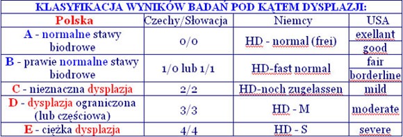
From January 1, 2001, the following symbols defining HD levels were introduced in Poland:

Progressive retinal atrophy – PRA

Note: the text was used on this website with the author's consent.
Any further distribution, commercial or otherwise, without the author's consent is prohibited.
In 1911, Magnuson first described progressive retinal atrophy (PRA) in a Gordon Setter dog. More in-depth studies of this condition in Irish setters were conducted by the English: Rasbridge, Hodgman, Parry and Lucas in the 1930s and 1940s. Research is constantly being conducted. Today it is known that the disease known as progressive retinal atrophy has various causes and different courses. It is expected that in the future many entities will be separated from this group of diseases. This disease is compared to retinitis pigmentosa in humans and is hereditary.
The classification of progressive retinal atrophy depends on the time of appearance of the first changes, the type of changes occurring in the retina and the mode of inheritance.
In some breeds, the first symptoms are noticed immediately after birth - changes in the fundus of the eye around the 10th week of life. In others, the first symptoms may appear in an adult dog.
The method of inheritance is also typical for the breed - most often recessive autosomal inheritance.
The basic symptoms include poor vision in the dark, which may develop into significant impairment of daytime vision or even complete blindness. As the disease progresses, we observe a decrease in the pupillary reaction and increased reflection from the fundus of the eye, often red in color. Examination of the fundus reveals excessive reflection of the fundus, a reduction in the number and narrowing of retinal vessels, and atrophy of the optic nerve papilla. Based on the interview and clinical examination, the presence of the disease can only be suspected. The final diagnosis is made after performing an electroretinography (ERG) test - which involves examining the electrical potentials generated when the retina responds to a light stimulus. The stimulus consists of precisely defined flashes of white light of varying intensity, as well as blue and red light. Based on this test, retinal dysfunction is diagnosed. In many countries, this is a mandatory test for breeding dogs belonging to breeds at risk of this disease.

Progressive retinal atrophy was diagnosed in dogs referred for consultation due to suspected eye disease as well as in dogs that came to the office for another reason, e.g. in puppies before the first vaccination. Routine clinical examination of puppies includes a shortened visual examination, including examination of the fundus of the eye. This makes it possible to recognize early symptoms of the disease. The observations described here concerned young dogs aged from a few weeks to 3 years. Out of 180 examined dogs, 32 cases showed clinical changes characteristic of progressive retinal atrophy. ERG was not performed in these dogs.
Typical symptoms were found in particularly at-risk breeds, such as long-haired miniature dachshunds, golden retrievers, setters, collies, and miniature schnauzers. The largest group were German Shepherds, several of which came from one breeding farm. This group also included hybrids.
The aim of my speech is to draw attention to the problem of hereditary eye diseases in dogs and to encourage people to deal with this problem following the example of other countries.

Ph.D. Marcin Pikiel
Specialized Veterinary Practice – Gdańsk.
e-mail

Granulomatous inflammation of the sebaceous glands – SA – (medical description)
Granulomatous sebaceous adenitis is a dermatosis characterized by progressive destruction of the sebaceous glands, most often found in young or middle-aged adult dogs, with breed predisposition: King Poodle, Visla, Akita, Samoyed, Terrier. The genetic basis for autosomal recessive transmission is suspected only in the King Poodle. In the United States, 25% in these dogs manifests subclinically.
There are various hypotheses regarding the etiopathogenesis, including: genetic destruction of the sebaceous gland, immunological destruction of the sebaceous gland, initial disorder of keratinization of the follicle causing clogging of the sebum ducts and destruction of the sebaceous glands, and anomalies in fat metabolism.
Clinical forms vary depending on the breed. Lesions in the King Poodle generally affect the facial area, forehead, earlobes, upper neck and trunk, as well as the tail. The lesions are characterized by partial symmetrical alopecia, excessive dandruff-like or psoriasis-like exfoliation, brittle hair, keratosis pilaris, rancid odor and variable itching.
The differential diagnosis includes demodicosis, dermatophytosis, and endocrine disorders. Histopathological examination of damaged tissue sections is diagnostic and shows granulomas with a predominance of lymphohistiocytes centered on the sebaceous glands and severe epidermal and follicular hyperkeratosis. In the case of old lesions, the absence of sebaceous glands, follicular fibrosis and atrophy of hair follicles are noted. These histopathological aspects are equally variable depending on the breed.

The therapy used varies depending on the type of lesions, their age and therapeutic approach. Here is a link on this website about homeopathic treatment of SA - in my opinion, the only one with good prospects. Because steroids are a dead end - as is often the case for such a primitive breed as the Akita - they simply shorten the dog's life instead of improving its quality.
Unfortunately, it is impossible to say what the basis of SA is. There is no evidence that the cause of this disease is genetic (however, as a precaution, dogs that have become ill should be withdrawn from breeding). So far, there is no way to check whether a given dog has SA until it becomes ill (and even then the result is often incorrect - see the link to "Kari's allergy story"). Therefore, not only in Poland but also in the world, there is no line free from this disease - in some apparently healthy dogs, the conditions necessary for the disease to manifest themselves simply did not occur. So if we want to eliminate SA completely, we simply have to stop breeding dogs. Primordial races are not adapted to the stresses of modern life in civilization, which life and man cope with increasingly worse. If any breeder boasts on his website that his breeding is free from SA, he is simply denying reality or simply hiding the truth. Personally, I am of the opinion that it is rather one of the diseases of civilization, because it usually occurs as a result of great stress and when the immune system is weakened (for example, after treatment with antibiotics). Akitas, as a breed with a narrow gene pool, are particularly susceptible to this type of diseases.


Babesiosis – a deadly disease transmitted by ticks!
Babesiosis is a disease transmitted by ticks that occurs in a large part of Poland. Forests and meadows, as well as parks and gardens covered with tall grass, are particularly at risk of ticks. The greatest number of ticks occurs in spring (April/May) and late summer (August/September), but they appear until snowfall.
Ticks attack dogs and cats as well as humans, and are extremely resistant (they can survive without a host for 3 years). Babesiosis is a deadly disease and difficult to treat, so it is particularly important to prevent it by regularly checking the dog's fur and removing any ticks, as well as using appropriate protective preparations. Treatment of babesiosis is expensive, much more expensive than the systematic use of preventive measures to protect against attacks by an infected tick. Symptoms of the disease may appear within a few days or a few weeks of infection. The development of the disease is often so rapid that despite immediate treatment, we can lose our dog within a few days.

The sucked tick introduces the pathogenic babesiosis protozoa (Babesia canis) into the dog's bloodstream, which attack red blood cells (erythrocytes), penetrating the inside of the attacked cell and tearing it apart (due to the rapid reproduction of the protozoa). Subsequently, subsequent red blood cells are attacked, which causes a chain reaction that destroys a significant part of them and causes ANEMIA. There is significant damage to internal organs, especially the kidneys and liver. Even though the kidneys have quite a significant reserve, their damage exceeding 75% makes it impossible for the animal to continue living. A partially damaged liver has the ability to regenerate on its own only to a certain extent. Additional effects of babesiosis may include severe circulatory system disorders and jaundice.

Characteristic symptoms of babesiosis:
apathy and lack of appetite - these are the first symptoms that are often ignored (the dog does not behave as usual, is lethargic and does not want to eat)
later, high fever appears (reaching 40-41°C) as well as vomiting and diarrhea (often bloody),
enlarged lymph nodes and yellowing of the oral mucosa,
problems with urination, hematuria (brown-colored urine),
respiratory and circulatory failure, nervous system disorders.
There is currently no vaccine against babesiosis available in Poland. A vaccine called Pirodog (by Merial) is registered in France and Switzerland.

Prevention involves using appropriate protective preparations that are absorbed through the animal's skin, preventing ticks from penetrating it and injecting infected blood. Although a tick can be found on an animal, it should not burrow into its skin or cause an infection. It will not enter your dog's bloodstream and will fall off within 48 hours. The effect of the preparations is limited (e.g. "Frontline" works on ticks for a month from the date of instillation on the skin - note, after using the preparation, you must not bathe your dog for 2 days), so you need to reuse it regularly.
Prepared by: Izyda Bach-Żelewska

Scientific study of granulomatous sebaceous adenitis
sponsored by JKC and WUAC at the University of Göttingen and a meeting of the WUAC Management Board
on October 19 and 20, 2004
On October 19 and 20, 2004, a meeting of the WUAC management board was held in Göttingen.
The WUAC website and the publication of a video about Akita dogs were on the agenda.
In the meantime, JKC has given WUAC its consent to publish a website, the current members of the WUAC board are of the opinion that the creation of such a WUAC website is necessary. At the very beginning, Fritz Paier from Austria volunteered to develop it and take care of this website as the material arrived.
I am full of hope that this website, at least in the German version, will be available by January 1, 2005 at the latest. http://www.wuac.info !
The mentioned JKC-Akita-video is partially made, it contains many interesting points. At the same time, many new ideas and wishes of management board members were discussed. For this reason, it is unlikely that this project (currently scheduled for English and Japanese) will be completed before mid-2005.
From my point of view, granulomatous sebaceous adenitis was the most important item on the agenda. That's why I'm a little disappointed that I can't write today that the research project will be continued.

The president of WUAC, Mr. Hoshi, was forced to cancel his participation due to an accident he suffered 5 days before the meeting, and Mr. Kamisato was also forced to stay in Japan due to his duties.

As a result, I was forced, as vice president, to chair the meeting, but of course I was not authorized to make financial decisions.

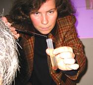
JKC assured that this decision would be made by the end of November. I hope that this decision will be positive for this project, because the facts that Dr. What Pfeiffer (pictured) and her team discovered are nothing short of extraordinary!

Here, I would like to thank all Akita owners who have so far supported this project and contributed to the health of the Akita breed and, I hope, will continue to do so!

The University of Göttingen is meanwhile collaborating with a number of scientists from around the world. The intensive exchange of information and ideas between scientists, new technologies as well as the luck necessary in scientific research have led to the fact that Göttingen is able to produce - after only ten months of work - extraordinary results.

Dr. Pfeiffer and her team found that both biopsies and blood from sick and healthy Akitas showed differences. Further biopsies and blood tests from Akita patients are urgently necessary to confirm these results.
By the way, a method of extracting RNA from blood was discovered for the first time. Until now, this method was completely unknown in molecular biology, hence it is a novelty on a global scale!

It seems that the University of Göttingen has discovered the key to deciphering granulomatous sebaceous adenitis through research so far and has found the right track to discovering why the cells of sick Akitas are going so crazy.
It was said very nicely at the conference: "There are many obstacles to overcome before a marker is found to identify carriers of this disease, but Mount Everest has already been conquered by Göttingen."
These were two exhausting days, filled with information and intense discussions. But also two days that brought a glimmer of hope that we will reach a point where we will be able to eradicate this terrible disease from this breed forever.
If this were possible for Akitas, it would mean great hope for the 44 other dog breeds in which the disease has also been diagnosed.
I can say one thing: I bow to the achievements and enormous commitment of the Göttingen Team as well as to the commitment of some Akita owners!
I must once again appeal to all owners and breeders of Akitas whose dogs have fallen ill - do not close your eyes to this disease, work actively with us, support research in Göttingen for the future of Akitas free from granulomatous sebaceous adenitis!

In this mood, I wish you all a Merry Christmas and lots of happiness and contentment in 2005. A great Christmas gift for all Akita owners would be an "OK" from JKC to continue funding this project!
Angelika Kammerscheid-Lammers
Please send biopsies and blood of Akitas suffering from granulomatous sebaceous adenitis to:
Dr. Ina Pfeiffer, Universitaet Goettingen,
Groner Landstraße 2, 37073 Goettingen
Arrangement of dates:
Telephone: 0049/551-39-9695 or 0049/551-39-3395.
Email: ipfeiff@gwdg.de
Every veterinarian has the necessary containers for two biopaths (from each dog) and blood test tubes (2 ml of EDTA whole blood). Biopathies are collected under local anesthesia.
If you have any questions, Dr. Pfeiffer is happy to be available by phone or email.
All data, unless expressly requested otherwise by the owner, are automatically treated as confidential.
translation from German: Darius Pollok

Homeopathic treatment of granulomatous sebaceous adenitis (effect photos)
Note: both text and photos used on this website with the author's consent.
Any further distribution, commercial or otherwise, without the author's consent is prohibited.

Below, Michael Rampak - the owner of an Akita named Bushi - born in June 1995, describes his own therapy, which has been effective for over a year. Bushie's problems with skin and fur began in September 1996. All possible tests and allergy tests were performed because there was no point of contact. In May 1998 Dr. Reichler from Zurich diagnosed granulomatous inflammation of the sebaceous glands.

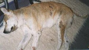
Bushi sick – April 1999, © 2000 M.Rampak

I only started thinking about putting him to sleep when I had already spent many thousands of marks on medications and therapies, and Bushie's condition was slowly but surely deteriorating. Today, when I see how, with relatively little effort, this terribly smelly, inflamed and hairless sneaking "something" became an Akita again, I can only be glad that I didn't pursue this thought to the end...

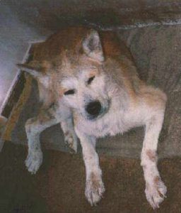
Bushi seriously ill in May 1999, © 2000 M.Rampak
STARTING THERAPY:
The last weekend of June 1999. All inflammations on Bushi's body were lubricated with calendula ointment (Calendumed Salbe). This softens the scabs and can then be removed painlessly for the dog after 4-6 hours. Where hair remnants are stuck to scabs, you can try to gently remove them using a lice comb. If you cannot remove them all at once the first time, please repeat the entire operation. In any case, scabs must be removed.
The dog should not be stressed, it must also have a break during these treatments. The next day and the next, we continue the treatment and continue to lubricate the inflammation 3-4 times a day.
Additionally, he received Bushi 250 mg of vitamin E, capsule daily, 3 x 5-10 Calendula balls (homeopathic medicine from marigold) D2 (drug concentration level), after 4 weeks D4 (drug concentration level). Simply pour a small spoon onto your dog's tongue.
A day or two later, when all the scabs have fallen off, oil the dog with St. John's wort oil (Hettral Johanniskrautöl). 100ml to 200ml - as much as needed, taking into account the condition of the rest of the coat, from head to paws, the dog must look as if it had fallen into a barrel of oil. It is important that the oil reaches the skin and then massage it in lightly. Be careful, not into the eyes or inside the ears. If the auricles are still dry, lightly massage the calendula ointment (Calendumed).
After oiling, keep your dog occupied for 2 hours, preferably go for a long walk, which will prevent the dog from licking the oil. Of course, a walk is only possible when it is warm outside. In winter, you need to spend these two hours in the bathroom with your dog and keep him occupied. In extreme cases, you can put a collar on the dog. After two hours, the dog should be bathed, for Bushi I use "Editerm" (by Virbac). Once a preliminary wash and then a proper bath. Do not rub when bathing, the oil should only be removed from the fur. Finally, rinse with "Humilac" (also by Virbac). After drying, the coat will still be a little oily, wipe it lightly with a paper towel from time to time. After two or three days, the remaining oil will be absorbed by the skin.
You can also make St. John's wort oil (Johanniskrautöl) yourself by mixing: 200 ml of high-quality olive oil and 10 ml St. John's wort tincture (Hypericum Urtinktur).
REPEAT THERAPY EVERY 10 TO 14 DAYS:
Repeat oiling and bathing after 10 to 14 days. After 8 to 10 days, Bushi developed large amounts of exfoliated skin and scratched himself very often during the first few days. At first I thought I had done something wrong, but it turned out to be a natural itch in the healing process.
The first thing that struck me after a few days was that the terrible smell that accompanied Bushi had disappeared. Now it smelled slightly of oil, and the skin on the hairless places that had first peeled off looked healthy.
Another 14 days later, I repeated the whole thing with baby oil (Bübchen), without any preservatives and perfumes. 200 ml of baby oil and 5 ml of calendula tincture (Calendula Urtinktur), leave for an hour or two and then, as previously described, bathe using a rinse.

FIRST SUCCESS AFTER 4 TO 6 WEEKS:
After 4 to 6 weeks, all of Bushie's superficial inflammation had healed and disappeared. The first puff appeared on previously bald spots.
Every 14 days, oil it, leave it for two hours, bathe and rinse. In mid-August, it looked like Bushie's hair exploded, all inflammation disappeared and he became active in a way I have never seen before. At the same time, his appetite increased and since I know that the fur consists largely of proteins, he received an additional portion of food every day.

Bushi's therapy brought results, September 1999, © 2000 M.Rampak

At the end of September 1999, the 85% achieved its former appearance. I extended the intervals between "oiling" Bushie to 3 weeks, now it is 4 weeks.
At the beginning of October, "OMNIFLORA N" was added to restore the intestinal flora, which had suffered due to many antibiotics.

Bushi, November 1999, © 2000 M.Rampak
I tried stretching it out to 5 or 6 weeks, but then my skin became dry and flaky again, so I quickly went back to the routine 4-week interval. Little has changed in the therapy, every four weeks apply baby oil, wait two hours, wash twice with "Etiderm" and rinse with "Humilac", eight caps per 1.5 liters of water. I also have to bathe him three times from time to time if I overdo it with "oiling".

I stopped taking the homeopathic medicine Calendula D 4 in January and he now only receives 250 mg of vitamin E twice a week. Since the beginning of therapy in June 1999, Bushi has not had any further inflammations and the coat is also quite fine, about 85 - 90 % of its previous appearance. At the beginning of the year he shed a lot and lost almost all of his undercoat, but it all grew back. In January I vaccinated Bushie, even though I had great doubts because every vaccine affects the immune system, but everything went well and in the future I will be able to vaccinate him quite normally.
What of these measures worked and what didn't - I cannot say yet. I think the decisive factor was St. John's wort oil (Johanniskrautöl). The result of the therapy lies next to me and smells of a dog - for me the most beautiful Akita in the world.
Michael Rampak (unfortunately the author is no longer alive)

PRODUCT INFORMATION:
Calendumed Salbe (Calendula ointment):
Deutsche Homöopathie-Union
DHU-Arzneimittel GmbH & Co. KG
Ottostr. 24
D 76227 Karlsruhe
tel.: 0721-4093-198 (Mon-Thurs.: 8.30 a.m.-4.30 p.m., Fri.: 8.30 a.m.-12.30 p.m.)
fax: 0721-4093-263; mailto:info@dhu.de
Hettral Johanniskrautöl (St. John's wort oil):
www.hetterich.de/Chemische/Produkte/Johanniskrautoel.html
Editerm and Humilac with Virbac:
Address of the international representative office of VIRBAC-Vertretungen
www.virbac.at/kontakt-int.html
Bübchen Babyöl (baby oil)
Omniflora N:
Novartis Consumer Health
www.novartis.de/novartis/html/d/consheal/otc/profil.htm

"Kari's Allergy Story"
Note: both text and photos used on this website with the author's consent.
Any further distribution, commercial or otherwise, without the author's consent is prohibited.

The little one was born on April 19, 2001 as one of eight in the litter. It comes from a small farm with extremely high standards. The breeder does not do it for financial reasons, he is an absolute lover of these dogs. The dogs were born and raised for the first weeks in conditions that many people, even in Europe, could only dream of. His breeding dogs (he also has "retired" dogs) were health-checked several generations ago, and everything was fine. I immediately liked the breeder; Before we were invited to meet the puppies' mother (they were five weeks old at the time), we were examined in an extremely pleasant conversation about our home conditions. It was done in an extremely charming and delicate way. Only after about an hour of conversation did we meet the puppies' mother. She ran to each of us (there were three of us - the whole family) and said hello. The dog was friendly and extremely interested in us. Only later did we find out that if the dogs' mother had not accepted us, we would not have "got" the puppy. And then, it was something special to mess with those eight balls of energy. I picked up our female dog, named Kari (Japanese for "hunt" or "wild goose" :-)) at the absolutely legal age of eight weeks and one day. I am writing about this in detail to emphasize that the breeder is an absolutely trustworthy and extremely responsible person, his dogs are genetically healthy, and my female dog's disease is only an individual problem. Thanks to his kindness, I was able to authoritatively check Kari's ancestors (in some cases up to great-grandparents) and siblings in the family tree. All conversations with the owners proved that the dogs from this breeding farm had absolutely no genetic predispositions.
Our redhead, then still a brown, round ball of arrogance and indifference, had been scratching himself intensely since the first day of his stay with us. We then fed Bosch food according to the breeder's recommendations. Smarkula was as picky as a princess, so you had to ask her several times to make her want to eat. A few days after moving to us, the first visit to the vet, examination of the puppy, setting the date of the next vaccination. I noticed to the vet that the female dog was scratching extremely intensely; the answer was, “All puppies scratch.” This was actually the beginning of my anxiety. Kari was growing and making noise like a good puppy. Only this itch, this itch…. The dog was scratching more and more, I heard the vet's quote more and more often... At the age of 3.5 months, the first rash appeared on his belly. Many conversations, consultations, ointments. A common opinion - it reacts badly to food, switch to dry food from another company. Be sure to dry them, because they are produced according to extremely balanced recipes and contain all the necessary ingredients. Well, let's get to work! New food was brought, praised and offered to the dog. I won't forget that disgust on the puppy's face for a long time! I was hard, the dog was hungry, so he ate! At the age of four months, another rash appeared on her tummy, then on her inner thighs. The fur has also disappeared in these places. The skin turned a strange brown color, the fur did not want to grow. From that moment on, negative changes on the skin began to occur. Allergic symptoms appeared in more and more different places, practically all rear parts of the body except the back and tail were affected by disease symptoms.


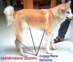
Losing fur, ugly, almost festering rash, severe skin redness. My trips to various veterinarians were longer and longer, and the circles in search of help became larger and larger. All blood tests, general dog tests and allergy tests were negative. The dog's condition was getting worse. Two more changes of food (according to the recommendations: "God forbid, give nothing other than dry food"), no improvement.
At the beginning of January this year, a skin biopsy was performed by a nearby veterinarian, with whom we had been "checking in" from the very beginning. This small piece of skin was to be examined at the pathology institute. After a week, we received a diagnosis that led us to nervous ruin: Sebadenitis! So far, the youngest case ever identified, atrophic sebaceous glands, there is no doubt...

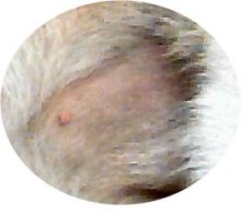
With such a diagnosis, one really loses the sense of reality, this dog has become an extremely important member of the family in the meantime! All documents and diagnoses collected so far were carefully sorted and sent by e-mail to Dr. Reichler in Zurich. After an earlier telephone conversation, she was kind enough to review all the documents and photos of the dog and express her opinion on the matter. Why Dr. Reichler in Zurich? – because she is the absolute authority in Europe (and probably in the world) in the field of granulomatous sebaceous adenitis. Mrs. Dr. Reichler ruled out granulomatous sebaceous adenitis with almost 100% certainty! On the one hand, a stone has fallen from our hearts, on the other - what is going on here??? What was I supposed to do? I called the pathology doctor who made the diagnosis. At first angry that a layman was asking her "absurd" questions, she was mollified and agreed to review all the test results and diagnoses again.


After all, this stubborn guy is a customer, he pays (and the bills are expensive), so he demands! After quite a long wait, the news came - don't worry, after reviewing all the documents and photos, it can be concluded that it is most likely not granulomatous inflammation of the sebaceous glands. Great, great, great - but what IS it???? The cycle of tests that Kari underwent in eight months exceeds the "human" average in a lifetime! All negative, the dog is actually healthy, the owner is probably a dog hypochondriac, and everyone who has seen the dog has visual hallucinations!!! There are a few things to understand about the story described here; the female dog was examined for all her organs, many people were involved and tried to help selflessly. Despite all efforts, the dog was in increasingly worse physical and mental condition.


There was no point in dreaming about educational work (let's not forget that this is an AKITA!!!) The prescribed "exclusion diets" did not lead to anything, the dog did not want to eat this crap. In mid-January this year, Kari went on strike - she stopped eating completely. She refused to eat completely, even eating bananas eagerly. She denied it. Me too, for five days. After five days, the dog was unsteady on its legs and the necessary walks were limited to meeting its physiological needs. It was clearly the beginning of the end. I gave up, I didn't care, as long as the dog started eating. I started cooking myself, after checking all the optimal possibilities according to the following recipe:
800 grams of lamb
2 kg of potatoes
2 kg of frozen vegetables (mixture).
No salt, no "flavor" additives.
At the beginning, this portion was enough for 1.5 days (the dog was emaciated and in poor condition), after two months the food doses were reduced so that this portion is enough for 2.5 days. Additionally, zinc is administered (the lack of zinc causes severe allergic symptoms, zinc is most often replaced by calcium, and the lack of zinc cannot be detected in blood tests). And because the diet is low in calcium, it is also given in tablets (theoretically, this contradicts the previous sentence, but in Kari's case it works perfectly).
As an additional sweet, only bananas, 2-4 times a week. Instead of dog "sweets", take 3-4 capsules a day with fish oil due to the omega acids and extremely high vitamin content. Bottled oil should not be used because the acids mentioned break down extremely quickly after opening the bottle and the agent becomes practically worthless.
In Kari's case, during the most intense itching attacks, short-term relief was provided by bathing in a special "Allercalm" shampoo (from Virbac), which moisturizes the skin. This shampoo is distributed only by veterinarians, all shampoos from specialist stores had the opposite effect - the dog went crazy with itching.
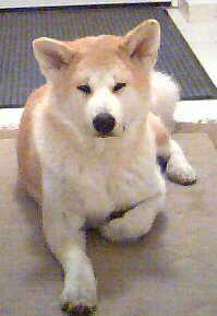
Why mutton? Because sheep are usually not inoculated with hormones and antibiotics. If there is no mutton, horse meat can be successfully used. Potatoes and vegetables as a carrier of vitamins and essential minerals, as well as to "fill the stomach". It is extremely important to make sure that the protein level in the food does not exceed 23%, otherwise the dog may "overfeed" with disastrous consequences. I will never forget the first time I cooked - Kari was sitting next to me in the kitchen and complaining loudly about the food. At that time, she was in a very bad condition - weak, physically poorly developed, small, always tired, without "seed", dull fur with large bald spots. She polished the first bowl of cooked food until it was shiny. The second one too, the third one…….After four extremely long weeks we noticed a clear improvement. The skin became better, allergic swelling became weaker and shorter. We continued to stick to the diet, all good advice was accepted with gratitude, carefully analyzed, and then most often ended up in the trash bin. Only what fit the concept was implemented. This week Kari will celebrate her first birthday and a happy birthday at that. She is almost healthy, there is no loss of fur, the itching is still there, but to a much lesser extent. The allergic swelling has almost completely disappeared; only in some places we can observe an unusually dark color of the skin (dark cherry), but without itching in these places. In the last three months, the female dog has grown an additional four centimeters, she is as strong as a Pittbull Terrier, cheerful and cheeky.
In general, all attempts at treatment with antibiotics, cortisone (fortunately sporadic) and ointments were without effect or with extremely short-term success.
The female dog is subjected to a thorough skin inspection every day and systematically observed. For now, the story continues to develop very positively, although the pace of improvement is not as rapid as at the beginning.

And the causes of the disease? Officially unknown, all test results were OK, no allergic reactions were detected. And yet... Personally, I am quite sure that the cause is dust mites. They are found on all dry-stored food, on rice, pasta, dog cookies (human cookies too :-)) ), bread. Cooking kills them, but the body reacts to their protein, regardless of whether they are alive or not. I was convinced of this by small "sins" such as offering the dog, for example, rice, a piece of roll, or dog cookies. Half a day later, the dog had no paws to scratch its entire body. An additional important factor, in my opinion, is high-quality food, which was able to improve the dog's immune system so much that the body began to defend itself. Only the future will show whether this will be possible with the 100%. For now, I will continue to follow the described diet until the symptoms disappear completely. Then I will begin to dilate it carefully, always calmly waiting for the dog's reaction.
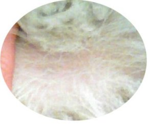
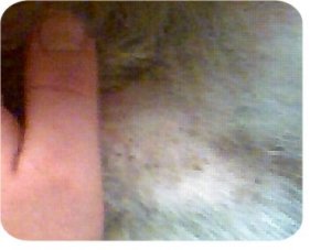
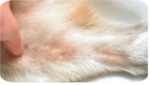
I am attaching photos of the female dog to this description. Photos with pasted text and clippings are from January 23 this year, approximately 10 days after starting the diet, the photo for comparison is from April 14 this year. Unfortunately, I lack photos from the time of severe disease, and in the photos presented, the skin condition was "softened" by the lens. In the final stage, the skin was in a disastrous condition, covered with an ugly, brown rash, and the dog was partially bald.
I described Kari's case quite thoroughly, but presenting all my mistakes when looking for the optimal diet would take even more time, strain the reader's patience and cloud the picture. However, if someone has similar health problems with their dog, they can try the diet just described. This way, the dog is spared a lot of suffering, vets have time to focus on other cases, and the dog owner's account remains normal 😉 .
I must emphasize one fact emphatically; I am not trying to question the knowledge and commitment of veterinarians in the slightest. Many people tried hard to help me, but due to the negative test results it was impossible for them. The conclusions I draw as to the causes of the disease are my own and only in Kari's case; the diet used is a combination of the experiences of veterinarians, dog owners, my observations of the dog and my own thoughts. Please do not treat this as a miracle cure for everything, I believe that in case of illness, it is absolutely necessary to consult a specialized veterinarian. Only if all else fails can you use the case described here as a basis for your own reflections.
I wish everyone healthy dogs

Dariusz Pollok (Germany)

Attention! The text was written by the author on April 17, 2002 especially for the website http://www.akita.pl/

Dermatoses of the nasal mirror
The nose mirror of carnivorous pets is a place particularly exposed to various injuries: mechanical or physical (UV rays), because it is not protected by fur and is the primary place of cognitive activity.
Anatomically, the nasal mirror is covered with thick epidermis, in which the lucidum layer is discreetly built up with compact orthokeratic keratin. In cats it is divided into round, knobby fields, and in dogs into small polygonal fields, which vary greatly between individuals and are considered the equivalent of human fingerprints. Although it is devoid of proper glands, the nasal mirror remains constantly moist thanks to the secretion of the serous glands, the lateral nasal gland and, additionally, many median nasal glands. In its anterior part, the nasal mirror is incised by a straight and deep groove. It has been experimentally confirmed that it is an indicator of the animal's good health, and any change noticed by the owner generally becomes a reason for a quick consultation with a veterinarian. Oculocutaneous syndrome
Vogt-Koyanagi-Harada Syndrome (VKH) The causes of VKH and the method of inheritance are not known (recessive gene hypothesis). VKH probably causes severe stress that weakens the dog's immunity... especially in combination with a vaccine - mainly against rabies, because this vaccine is very harmful to the puppy's body...
No breeding line is free from VKH - at most, the case has not yet emerged, has not been properly diagnosed, or the breeding is simply still in its infancy. It is not an infectious disease and is probably genetically determined. However, it is not possible to diagnose the disease before it manifests itself. The only thing that responsible breeders can do today is (despite the lack of regulatory requirements) regularly examine the eyes of their breeding dogs. If we wanted to withdraw from breeding the lines in which VKH occurred, we would have to stop breeding breeds in which this disease occurs. Fortunately, it is an extremely rare disease, which will certainly not comfort the owners of a dog who has been unlucky enough to suffer from it, as it is an extremely serious disease. Well, everyday stress causes more and more diseases in people, and dogs are even more exposed to it, because civilization destroys them faster than humans. If we want to reduce the likelihood of our dog getting sick, do not expose it to stressful situations, do not vaccinate against rabies too early, and before vaccination, make sure that the dog is in perfect condition. Let's make sure the vet thoroughly examines the puppy before vaccinating it. If he wants to vaccinate immediately without an examination, let's change the office... The disease attacks in stages.
Ocular stage -> acute conjunctivitis, tearing, scleritis, eyelid swelling, possibility of blood clots formation (they should be removed immediately). Affected eyes are extremely painful due to increased pressure inside the eye (bulging eye) and detachment of the retina (in some people, the eyeballs are even removed). A characteristic symptom is a milky-blue haze in the eyes. In many cases, glaucoma, cataracts, optic nerve atrophy, and complete/partial blindness occur.
Dermatological stage -> gray hair, hair loss, discoloration of: eyelids, nose, lips, palate, paw pads, scrotal area, vulva and anus. The entire body is rarely affected. Ulceration, difficult-to-heal crusts and erythema may occur in places of discoloration. The area around the skin lesions is itchy and painful.
Skin changes often occur simultaneously with the ocular stage or immediately after it. Not all of the above. symptoms must occur. The onset of the disease may be slow (hard to notice) or sudden. The degree of advancement and speed of development of the disease varies. Quick help and the use of appropriate medications can stop the development of the disease, but relapses may occur.
a) Epidemiology: very rare
Predisposed breeds: Akita, Samoyed, Chow-Chow, Siberian Husky, Irish Setter, Golden Retriever, Saint Bernard, Old English Sheepdog, Shetland Sheepdog, Australian Shepherd, Dachshund, Fox Terrier. The first symptoms of the syndrome most often appear in dogs aged 15 months to 2 years (exceptionally younger than 1 year or older than 4 years), and both dogs and female dogs are affected.
b) Clinic: Severe uveitis preceded by depigmentation of the mucous-cutaneous junctions of the muzzle.
c) Diagnostics: Histology: lichenifying intercellular dermatitis
Treatment:
– ophthalmological: subconjunctival administration of corticoid and mydriatic drugs.
– general corticosteroids + azathioprine (Immuran).

Stomach volvulus (flatulence)
In addition to Akitas, German Shepherds and Great Danes are also susceptible to this disease.

The digestive tract in large breed dogs accounts for only 2.7 % of the total body weight. If the amount of food consumed is too large, the dog's stomach may become twisted and enlarged. This causes pressure on the spleen, diaphragm and blood vessels (impediment to blood flow), which may lead to gastric torsion, commonly known as flatulence.
Initially, the animal salivates heavily, its stomach becomes hard and painful, and the dog constantly tries to vomit. Then he lies down, it swells quickly, and breathing problems occur. Failure to take any action may result in the animal's death due to circulatory and respiratory disorders. You should then go to a veterinarian immediately.

The problem is that the dog's stomach, shaped like a bellows, expands, turns and twists, trapping gas, food and water inside. The blood vessels twist, cutting off the flow of blood from the stomach and spleen to the heart. Blood cannot carry oxygen to these tissues, and poisonous products of cellular metabolism accumulate in them. Under their influence, the animal experiences shock. The patient's condition is rapidly deteriorating. Immediate help is needed, every minute counts. Surgery must be performed because without it it is impossible to detect damage to the stomach and spleen. The procedure is always risky, so you should choose a good doctor.
Procedure: the dog lies on the table with its belly up in the operating room. Its mouth is open and its tongue hangs to the side around the endotracheal tube. It is covered from head to paws with blue paper, only its belly is exposed. The surgeon cuts the abdomen. If the dog is lucky and both the liver, spleen and stomach are fine, the doctor attaches the stomach to the abdominal wall so that it does not twist again. The entire area is then rinsed with a saline solution.
The operation, performed by a good surgeon, should be successful...

Puppy's ears not erect - what to do
If your puppy's ears do not stand up, you should make sure that he is provided with the necessary ingredients in his food as soon as possible.
You should give him chicken feet - boiled or raw (clean and preferably scalded) - of course only after cutting off the claws. It is also recommended to feed large amounts of poultry and veal cartilage, as well as bone meal. Some people also serve pig ears and tracheas, but pork is generally not recommended for dogs.
You can also try gluing the ears so that they stand up and strengthen the cartilage. However, remember to allow air to enter the ear, otherwise it may result in a fungal infection.

error: Sorry, that function is disabled. Copyright 2000-2023 by Izyda Bach-Żelewska. Shouldn\\\\\\\'t be taken from this site.